Dog care services in Tonbridge
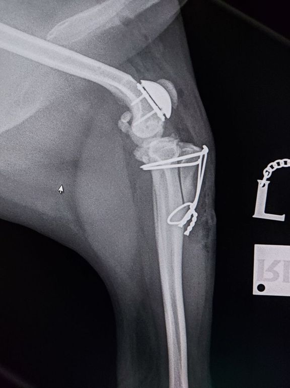
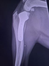
Patella Luxation repair with a prosthetic groove and tibial tuberosity transposition
Medial Patella Luxation
Medial Patellar Luxation (dislocating kneecap) is a common developmental abnormality in dogs, especially small and medium sized breeds. This can cause varying degrees of lameness and development of osteoarthritis over time.
We perform the latest techniques most suited to ensure the best chance of recovery including RidgeStop™ Prosthesis, Tibial Tuberosity Transposition, Recession Sulcoplasty, Femoral Varus Osteotomy and Patella Groove Replacement.
This sore patient has been suffering with grade 4 medial patella luxation on both legs.
In this severe case, the patient underwent Patella Groove Replacement where the part of the bone with the groove that the patella is supposed to slide in up-and-down has been cut-off and replaced by a prosthetic artificial one. Additionally, part of the tibia (shin bone) has been surgically repositioned in order to re-align the patella with the new artificial groove (called a Tibial Tuberosity Transposition).
Within 10 days, this poorly doggy was already using his bionic leg without any lameness or discomfort.
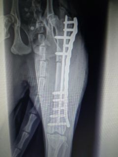
A femoral fracture repair with a locking plate and intramedullary pin
Fracture repairs
TPLO in a dog with a torn cruciate ligament
Cranial Cruciate Ligament Rupture


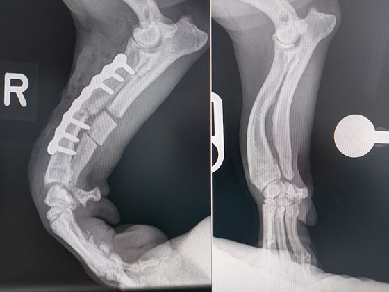
Angular Limb Growth Deformity
Angular Limb Growth Deformity is a developmental abnormality of the leg causing it to turn and twisting outwards, making it increasingly difficult to walk properly without knuckling over.
In this case, the deformity of the leg was corrected by cutting the bones in several places and rotating the bone segments by specified angles before securing it with a special locking metal plate and screws.
This procedure required preliminary xrays, a CT scan and meticulous planning on a computer in 3 dimensions to get the optimal angle for the bone cuts.
After the 3D planning on the computer was done, a 3D model of the leg before and after the correction was printed on a 3D printer, and special saw guides were 3D printed as well to enable a very accurate surgery.
The printed 3D model also enabled the special metal plate with the screws to be accurately planned and contoured exactly to the shape of the bone, so that during surgery it could be perfectly positioned.
Four days after surgery, this patient was already using the leg well without any knuckling anymore (still with a slight limp as expected, until the bone completely heals over the next 3 months).
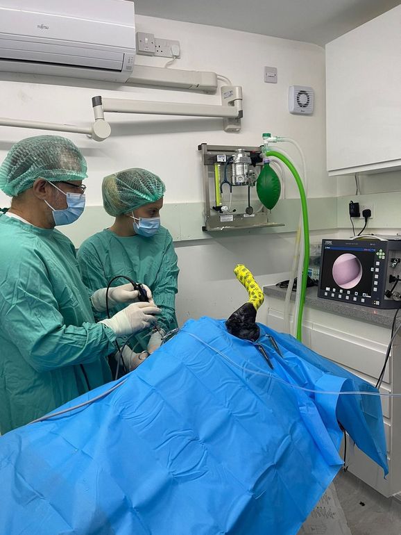
Tommy and Keri performing elbow arthroscopy (key-hole surgery) in the surgical theatre.
Arthroscopy
Here is a little behind-the-scenes sneak peak of veterinary surgeon Tommy and registered veterinary nurse Keri performing an arthroscopy (keyhole procedure) on both elbows of a lovely patient of ours. This poor doggy was quite lame as a result of elbow dysplasia - typically seen in Retriever breeds.
Arthroscopy involves the placement of the endoscopic instruments within a joint via keyhole incisions, allowing meticulous visualisation of the cartilage surfaces and ligaments.
Arthroscopy allows much more detailed evaluation of the joint compared to standard radiography (x-rays). Arthroscopic surgery has revolutionised the treatment of many orthopaedic conditions, such as meniscal injuries in the knee joint, elbow dysplasia and osteochondritis dissecans (OCD).
Endoscopy and endosurgery
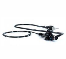
Gastroscopy