Dog care services in Tonbridge
Surgical intervention of cranial cruciate ligament disease using the Tibial Plateau Levelling Osteotomy (TPLO) technique.
Cranial Cruciate Ligament Disease
The cranial cruciate ligament (CrCL) in dogs (known as the “anterior” cruciate ligament in humans) is a band of fibrous tissue that attaches the upper thigh bone (femur) to the lower shin bone (tibia). Its function is to prevent the tibia from shifting forward relative to the femur when weight is applied, as well as preventing the stifle joint from over-extending or rotating.
Unfortunately, it is not uncommon for this ligament to weaken and fray over time, especially in medium/large breed dogs - Labradors, Rottweilers, and Boxers. As the structural support of the joint becomes loss, the femur has a tendency to slide and grind against the tibial surface resulting in a painful lameness and osteoarthritis.
We offer a range of surgical corrections such as Tibial Tuberosity Advancement (TTA), Tibial Plateau Levelling Osteotomy (TPLO), Modified Cranial Closing Wedge Osteotomy (MCCWO) and Lateral Suture.
The Tibial Plateau Levelling Osteotomy (TPLO) is the most robust surgical technique, and is advised in heavier dogs. This surgery involves creation of a radial cut in the top of the tibia and rotation of the segment until the slope in the bone is no longer present. The bone is subsequently fixed in this new position using a locking plate and screws.
The recovery period is approximately 6-8 weeks whilst the bone heals.
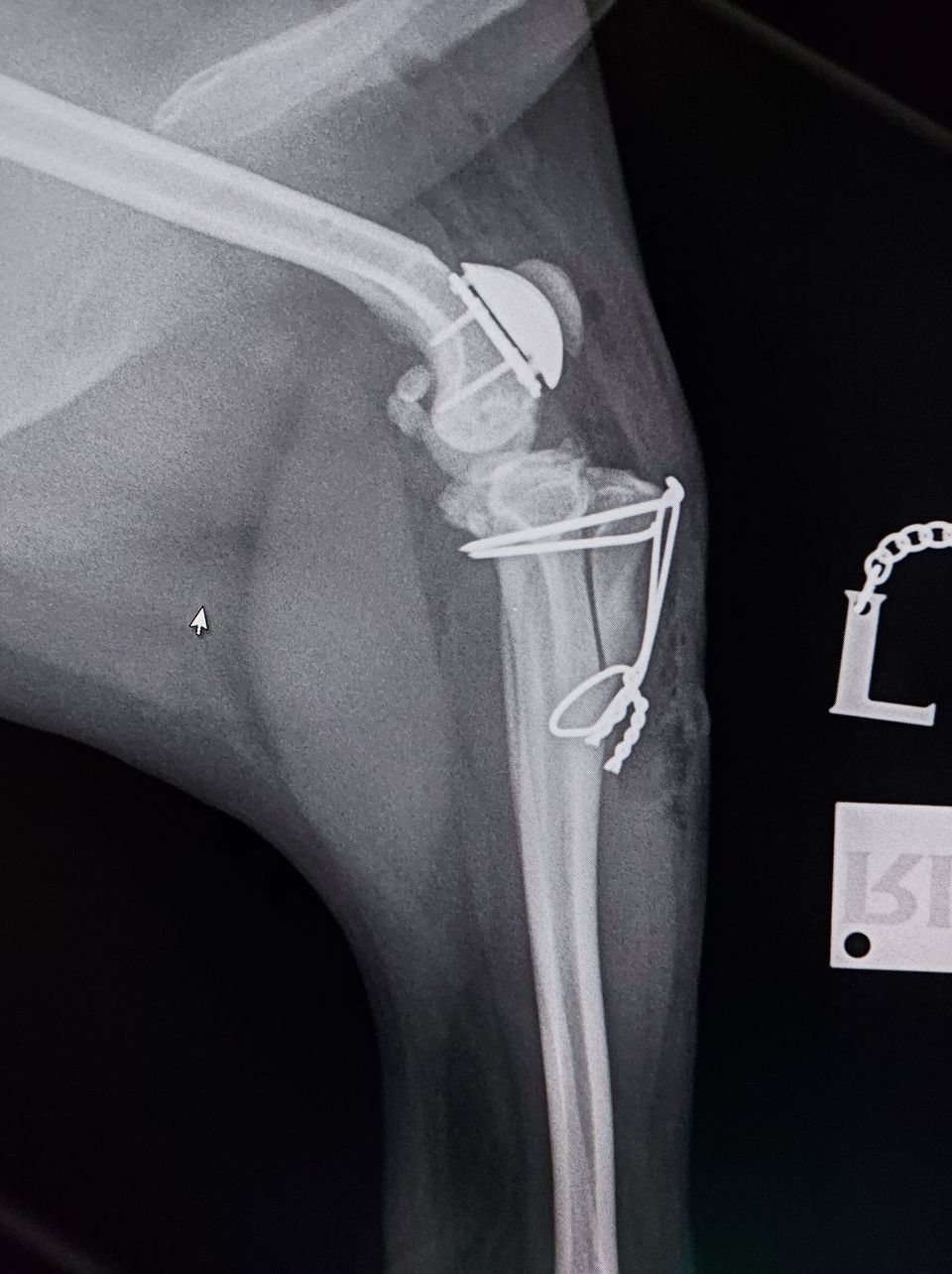
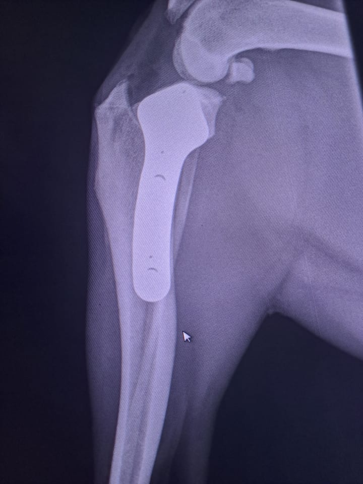
Patella Luxation repair with a prosthetic groove and tibial tuberosity transposition
Medial Patella Luxation
Medial Patellar Luxation (dislocating kneecap) is a common developmental abnormality in dogs, especially small and medium sized breeds. This can cause varying degrees of lameness and development of osteoarthritis over time.
We perform the latest techniques most suited to ensure the best chance of recovery including RidgeStop™ Prosthesis, Tibial Tuberosity Transposition, Recession Sulcoplasty, Femoral Varus Osteotomy and Patella Groove Replacement.
This sore patient has been suffering with grade 4 medial patella luxation on both legs.
In this severe case, the patient underwent Patella Groove Replacement where the part of the bone with the groove that the patella is supposed to slide in up-and-down has been cut-off and replaced by a prosthetic artificial one. Additionally, part of the tibia (shin bone) has been surgically repositioned in order to re-align the patella with the new artificial groove (called a Tibial Tuberosity Transposition).
Within 10 days, this poorly doggy was already using his bionic leg without any lameness or discomfort.


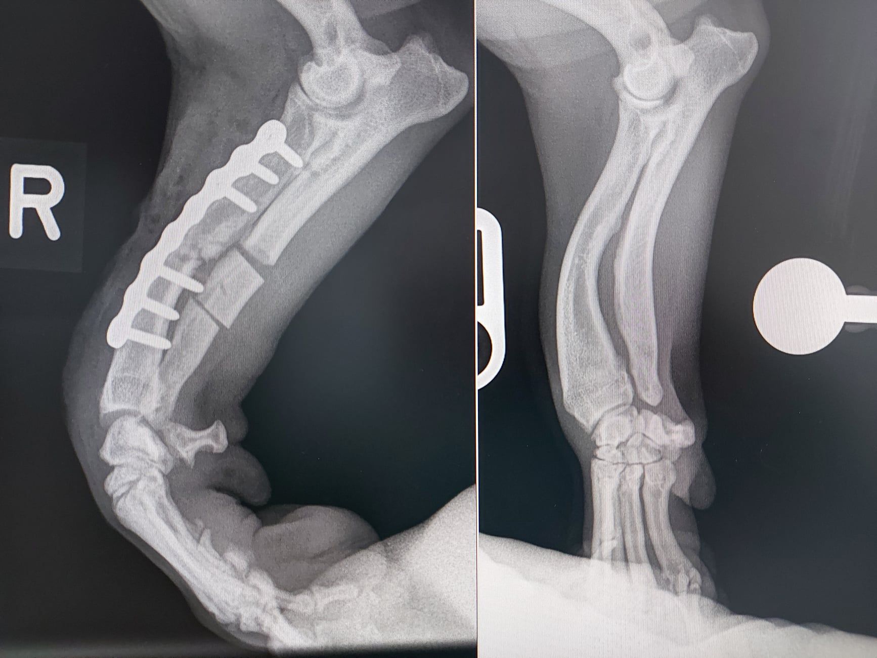
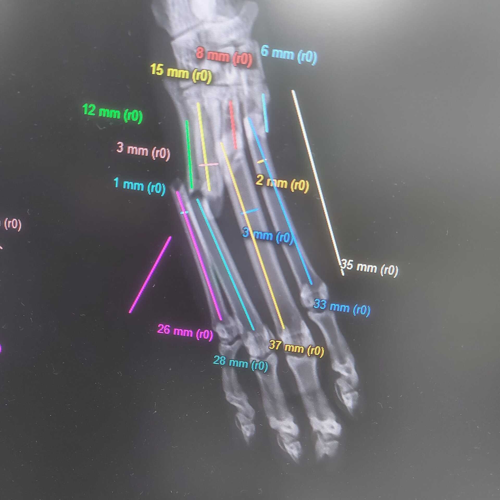
Angular Limb Growth Deformity
Angular Limb Growth Deformity is a developmental abnormality of the leg causing it to turn and twisting outwards, making it increasingly difficult to walk properly without knuckling over.
In this case, the deformity of the leg was corrected by cutting the bones in several places and rotating the bone segments by specified angles before securing it with a special locking metal plate and screws.
This procedure required preliminary x-rays, a CT scan and meticulous planning on a computer in 3 dimensions to get the optimal angle for the bone cuts.
After the 3D planning on the computer was done, a 3D model of the leg before and after the correction was printed on a 3D printer, and precise saw guides were 3D printed as well to enable a very accurate surgery.
The printed 3D model also enabled the special metal plate with the screws to be accurately planned and contoured exactly to the shape of the bone, so that during surgery it could be perfectly positioned.
Four days after surgery, this patient was already using the leg well without any knuckling anymore (still with a slight limp as expected, until the bone completely heals over the next 3 months).
We have extensive orthopaedic experience and expertise, and can repair broken bones using the latest technologies and techniques.
A full surgical team including the surgeon and nurses make sure the whole operation goes smoothly and safely.
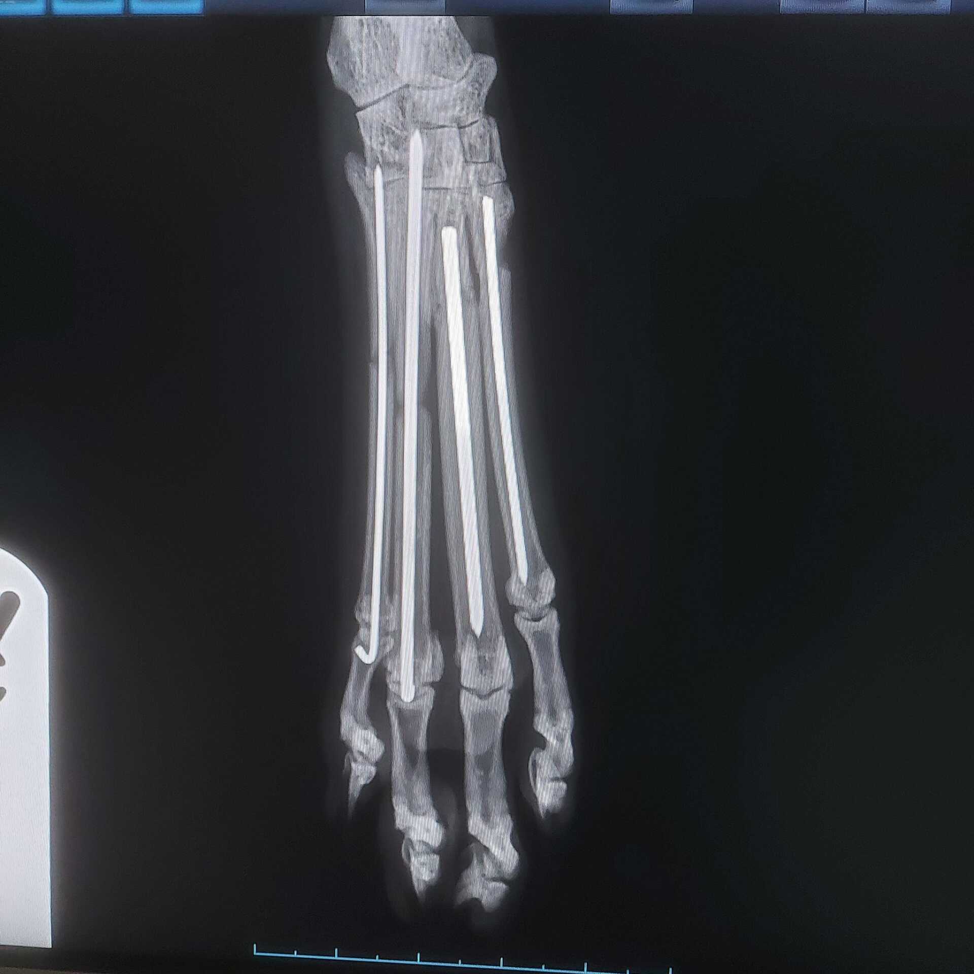
Multiple metatarsal fractures in a cat.
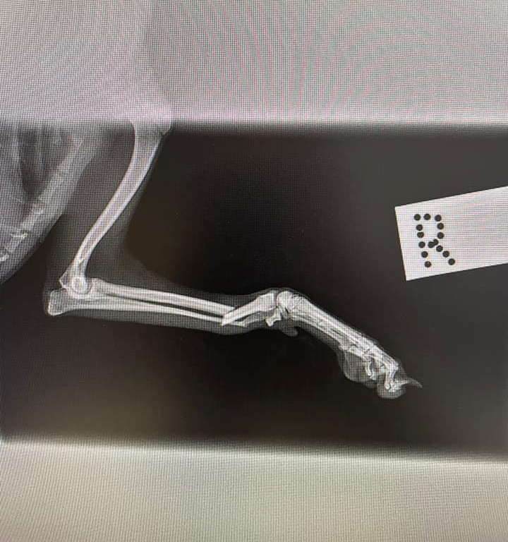
Distal radio-ulnar fracture in a toy breed dog.
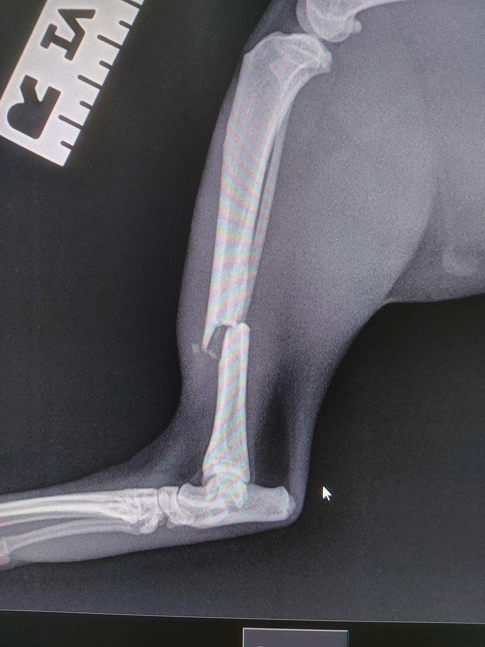
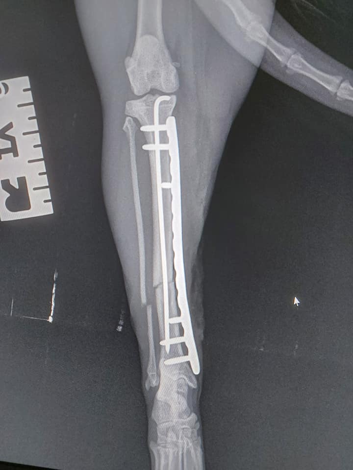
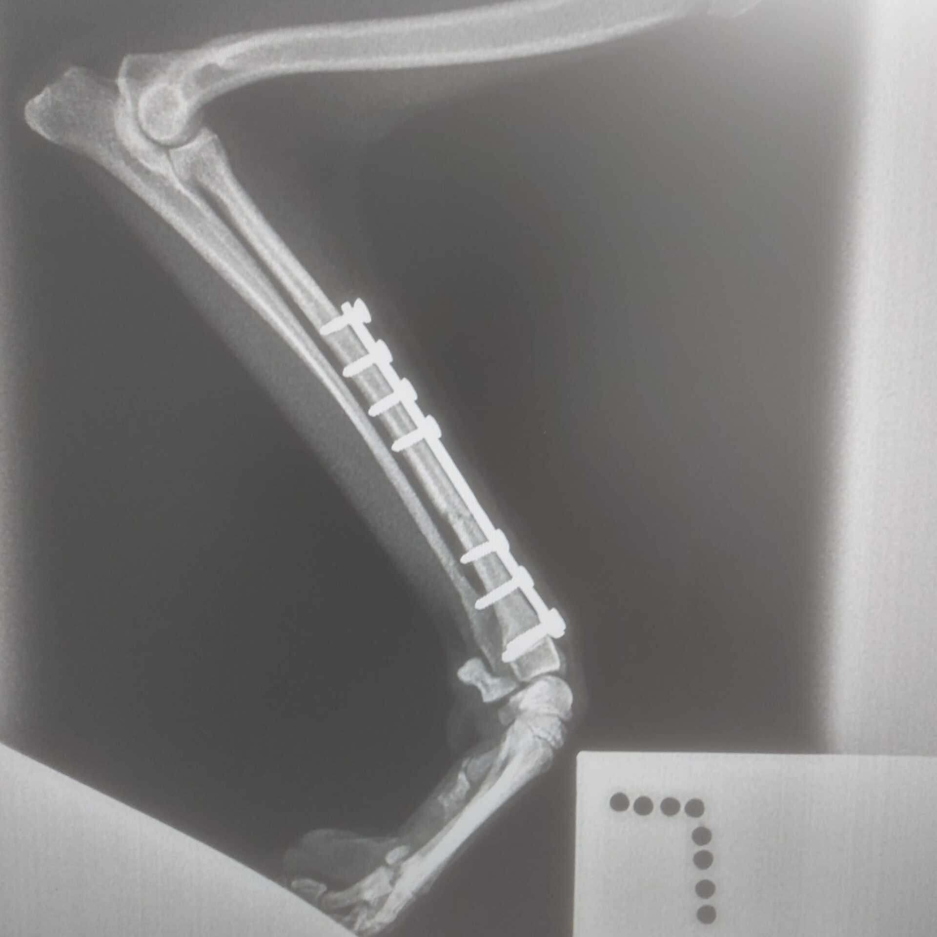
Transverse mid diaphysial fracture of the tibia and fibula in a cat - repaired with a classic pin/plate combination.
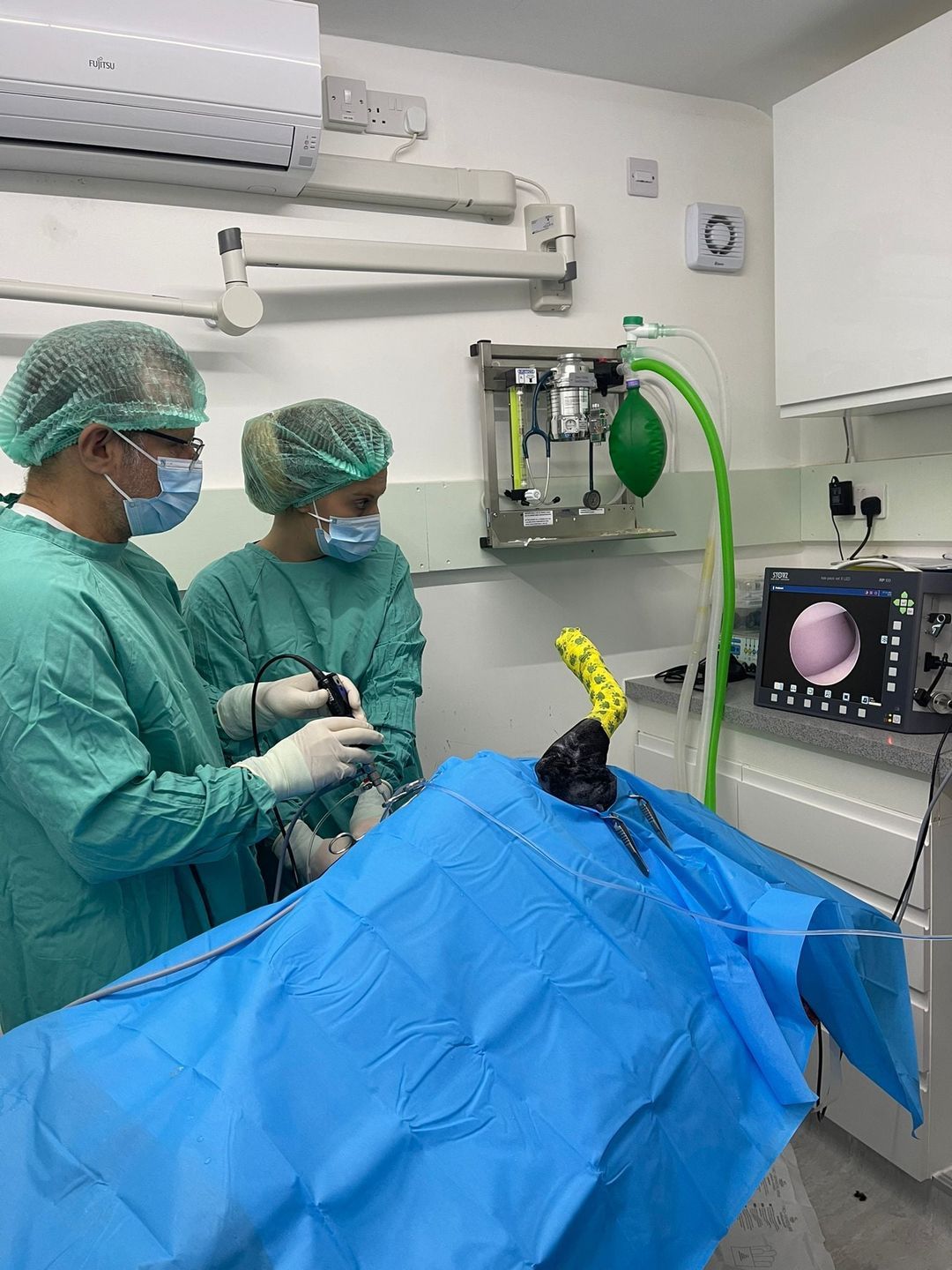
Tommy and Keri performing elbow arthroscopy (key-hole surgery) in the surgical theatre.
Arthroscopy
Here is a little behind-the-scenes sneak peak of veterinary surgeon Tommy and registered veterinary nurse Keri performing an arthroscopy (keyhole procedure) on both elbows of a lovely patient of ours. This poor doggy was quite lame as a result of elbow dysplasia - typically seen in Retriever breeds.
Arthroscopy involves the placement of the endoscopic instruments within a joint via keyhole incisions, allowing meticulous visualisation of the cartilage surfaces and ligaments.
Arthroscopy allows much more detailed evaluation of the joint compared to standard radiography (x-rays). Arthroscopic surgery has revolutionised the treatment of many orthopaedic conditions, such as meniscal injuries in the knee joint, elbow dysplasia and osteochondritis dissecans (OCD).